
By Chris Faubel, M.D. —
This is a PERFECT example of why interventional pain physicians should both consider getting an MRI before performing any epidural steroid injections and certain other procedures, and also to look at the images yourself. Also, make sure you look at both sagittal and axial images, as well as the T1 and T2 weighted images
Patient Presentation
Complaints consistent with a right L5 radiculopathy that started two months ago but has gotten much worse the past two weeks. No history of trauma or other inciting event.
MRI Findings
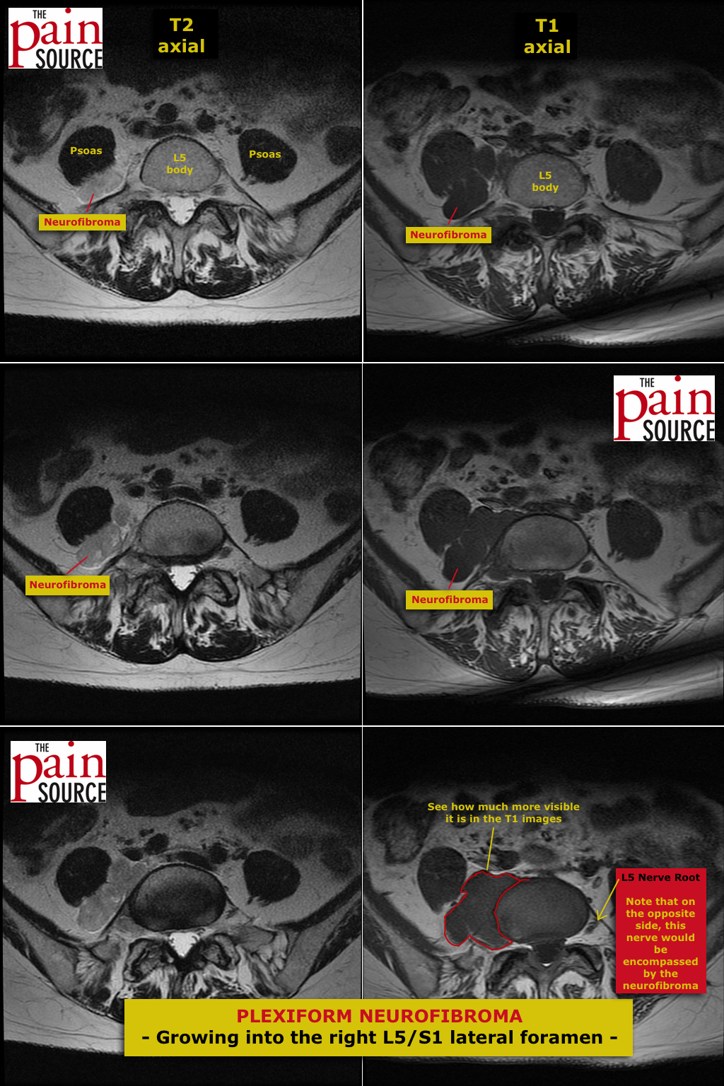
MRI shows severe right facet arthropathy, a mild right lateralizing disc bulge, and grade 1 spondylolisthesis all contributing to moderate right L5/S1 foraminal stenosis. This could explain the patient’s symptoms alone.
Surprise finding: Large plexiform neurofibroma growing into the right L5/S1 neuroforamen. Most visible on the T1 axial images; at a quick glance of the T2 axial images, this neurofibroma can possibly be missed.


")

