One video below. Click the black title link above (if you can’t see the video).
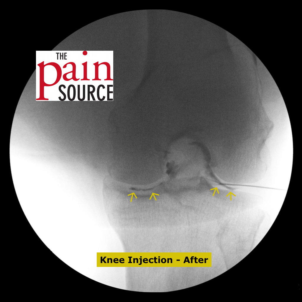
1) Left knee injection under fluoroscopy. It is important to note the “mustache sign” in which the contrast spreads to both sides of the joint on the A-P view. Also note the lateral fluoro view that shows contrast spread into the suprapatellar bursa — this is observed in 85% of adults as the septum becomes perforated during the 5th month of development. You will find fluid in the suprapatellar bursa with MRI and ultrasound in patients with knee joint effusion or bursitis. The image of the Baker’s cyst is great in that it shows that they are connected to the knee joint; they are especially common in patients with meniscal tears in which the knee has an effusion that leaks into the cyst and causes a fullness feeling in the back of the knee.





Hi. I am dr. Hanan from benghaZi. Libya. I ve master degree in physical medicine and rehabilitation since 2010. I interested in fluoroscopy injection. Where can I have training for it.
the Spine Intervention Society based out of the USA has international courses every year. I believe the website is spineintervention.org