By Chris Faubel, M.D. —
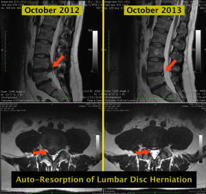
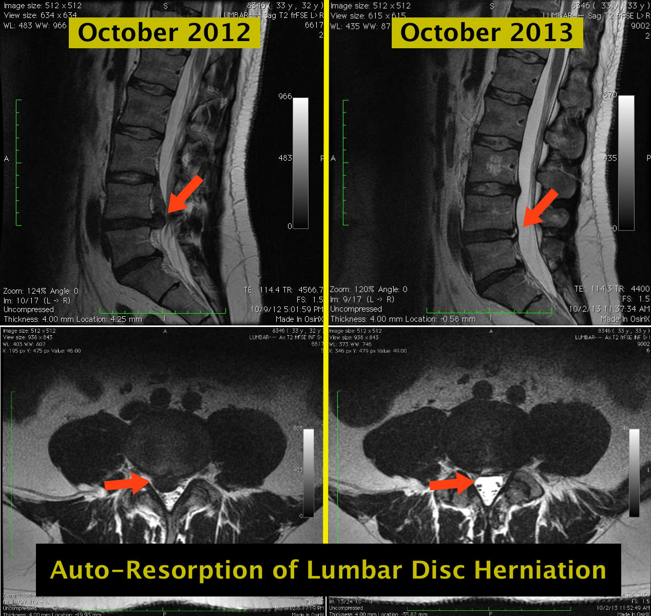
This is an excellent example of an MRI showing resorption of a rather large lumbar herniated disc (a year after the first image). It is rare to see such a dramatic before-and-after, because either the patient has surgical excision of a disc this size, or because the disc resorbs and they no longer have back pain so a follow-up MRI is never taken.
This particular patient (32-33 year old male) had both MRIs taken while admitted to a local hospital for severe low back pain. The patient didn’t have any neurologic deficits in October 2012, so no surgery was performed. A short course of oral steroids and some physical therapy resolved the low back pain. Then, in October 2013, the severe pain returned and another MRI was taken.
This spontaneous, dramatic auto-resorption of the large disc herniation is a great patient education tool for patients with these imaging findings. “Yes, your disc herniation can just go away by itself sometimes.”




It is a beautiful example of resolution of the HNP but you make no mention of the annular tear, quite possibly the source of his continued pain.
That’s quite possible. The post was to highlight auto-resorption of a lumbar disc herniation. Not really to discuss the patient’s lumbar pain at the time of the second MRI. But yes, possibly that annular tear. Good job Michael
Yes, look at this article too, available in the British Medical Journal Case Reports, 2013 Sep 30.
Available at:
https://www.researchgate.net/publication/257249779_Natural_resolution_of_a_herniated_lumbar_disc
or
http://casereports.bmj.com/content/2013/bcr-2013-201037.long
This is an extremely common phenomenon that few know about, including physicians. Thanks for posting.
Thanks Fred