By Chris Faubel, M.D. —
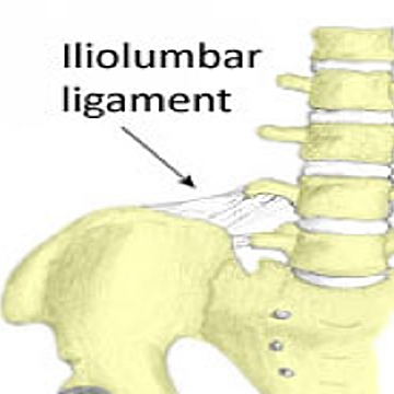
To learn about Iliolumbar Syndrome, follow this link.
ICD 10 code: M46.06 (lumbar spine enthesopathy)
CPT codes:
- 20550 “Injection(s); single tendon sheath, or ligament, aponeurosis”
- 77002 “Fluoroscopic guidance for all types of needle placement, i.e., biopsy, aspiration, injection, or localization device”
PROCEDURE TECHNIQUE:
Solution: Varies depending on the number of sites that you plan to inject. Typically, I inject four sites and use a 4-ml solution consisting of 3-ml of 0.5% bupivicaine and 1-ml of Depo-Medrol 40mg/ml. You can also use just local anesthetic if the patient has an elevated blood sugar.
Position: Prone
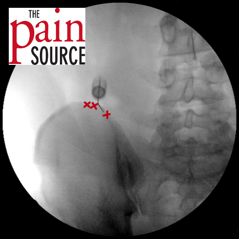
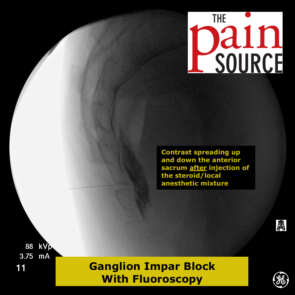
Fluoroscopy: An 18-gauge 1.5″ needle tip is placed on the cleaned skin over the patient’s point of maximal tenderness along the iliac crest. An A-P fluoroscopic view is used to visualize the iliac crest under this needle.
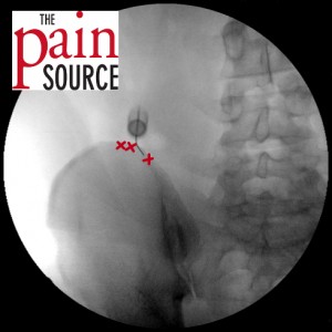
Technique: First, create a skin wheal and anesthetize the deeper subcutaneous skin with 1% lidocaine and a 25-gauge 1.5-inch needle. Next, a 22- or 25-gauge Quincke needle is used to advance down to the area of the posterior edge of the iliac crest at the point of maximal tenderness. Inject 1-ml of the solution here. Then, pick three or four other sites on either side of the initial injection site (see the picture to the right).
Expectations: Patient should have significant reduction in pain within a few minutes if this is the real source of his/her pain; this injection is then both diagnostic and therapeutic.






Would you code 20550 for interspinous ligament injections as well and add 59 for each level?
Thank you!