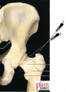
Technique: Lateral approach with fluoroscopic guidance. Note: I GREATLY prefer the A-P approach. Check it out HERE.
Outpatient procedure
Patient position: Prone
Needle used: a 22-gauge 3.5″ quincke spinal needle is sufficient for most patients, although a 5″ may be needed for larger patients
Steps:
- Prep and drape
- Use the fluoroscope to find the skin over the greater trochanter
- Make skin wheal and deeper anesthesia with local anesthetic (1% lidocaine) with 27- or 25-gauge needle at a spot 4-6cm cephalad to the greater trochanter
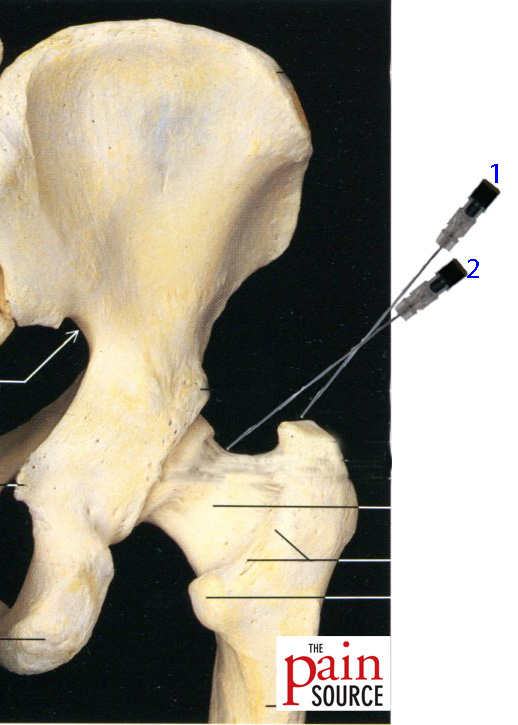
- With 3.5″ 22-gauge needle, enter the skin through the skin wheal and go towards the top of the greater trochanter (#1in the photo)
- This will tell you you’re in the correct coronal plane
- Redirect the needle and aim towards the femoral head/neck junction (#2in the photo)
- Do NOT try to inject between the femoral head and the acetabulum
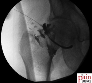
- Inject contrast to confirm intraarticular flow
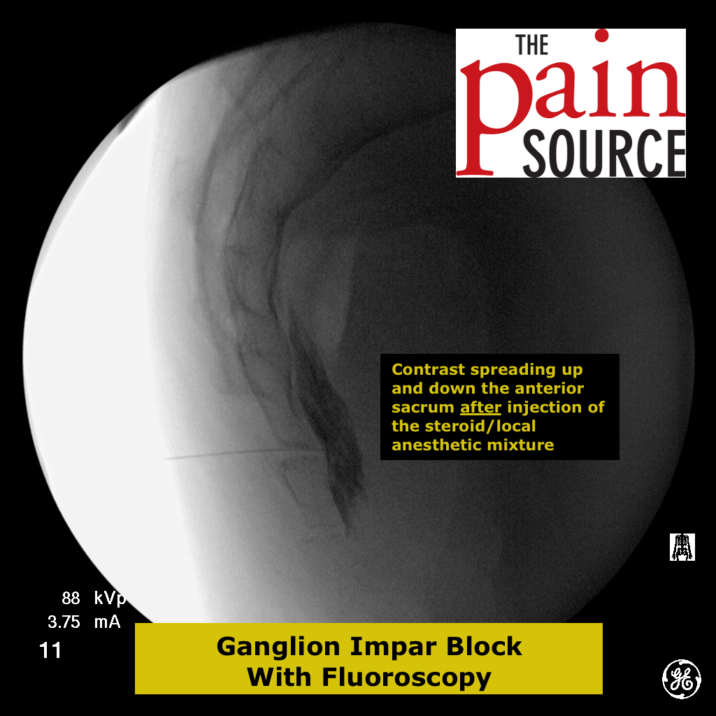
- You should be looking for the circular pattern noted in the fluoroscopic image below
- Inject the corticosteroid or steroid/local anesthetic injectate
- Remove needle. Place band-aid. Patient can go home immediately, with instructions to take it easy with that hip for 3-5 days.





Nice work!
Is there a benefit of going anterior vs lateral approach?
@Azlan:
http://www.ncbi.nlm.nih.gov/pubmed/11603669
Here’s a study in which they injected 15 cadavers (30 hips) with a lateral approach on one side, and an anterior approach on the other. They did NOT use fluoroscopy. They kept the needles in and dissected each cadaver to see whether the dye was intraarticular with either approach, and whether the needles were piercing or near any neurovascular structures.
The lateral approach was successful 80% of the time, and the needle never came within 25mm of any neurovascular structures.
The anterior approach was successful 60% of the time, but was piercing or in contact with the femoral nerve 27% of the time, and within 5mm of the nerve 60% of the time.
Commentary: We must note this is a cadaver study, but that the risk to neurovascular structures is still much more with an anterior approach. But to answer your question, I’d presume the efficacy would be the same as long as you injected the steroid into the joint.
Thank you. That was very helpful.
why not to inject between the femoral head and the acetabulum?
Good question. For one, the actual femoral head is articulation with the acetabulum is medial enough that you’re more likely the contact the femoral artery as you go down. Also, you don’t want to potentially damage the articular surface. It’s much easier to just get the needle tip under the large hip capsule.
Are patients usually under light sedation for this procedure?
In my opinion and that of the Spine Intervention Society, IV sedation doesn’t need to be used for this procedure. Local anesthesia (lidocaine) is all that’s needed.