By Chris Faubel, MD —
J Bone Joint Surg Am. 2001 Sep;83-A(9):1306-11.
The value of magnetic resonance imaging of the lumbar spine to predict low-back pain in asymptomatic subjects : a seven-year follow-up study.
Borenstein DG, O’Mara JW Jr, Boden SD, Lauerman WC, Jacobson A, Platenberg C, Schellinger D, Wiesel SW.
Division of Rheumatology, George Washington University Medical Center, Washington, DC 20037, USA.
http://www.ncbi.nlm.nih.gov/pubmed/11568190
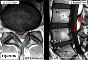
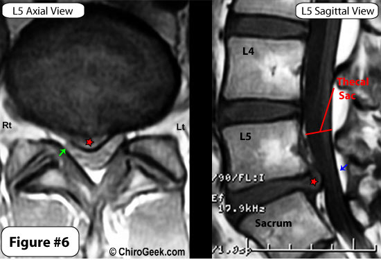
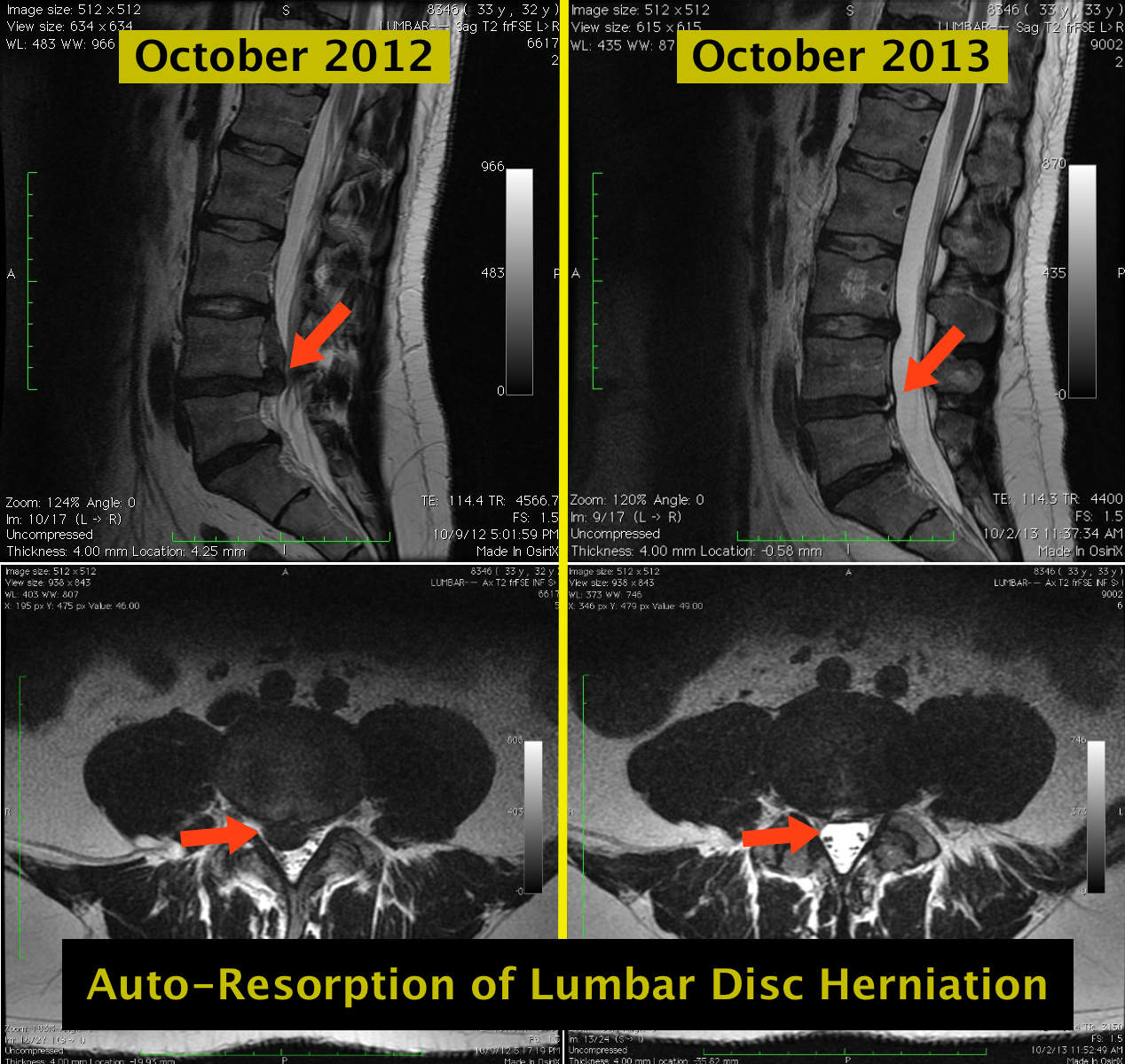
Photo courtesy ChiroGeek.com
ABSTRACT
BACKGROUND: In 1989, a group of sixty-seven asymptomatic individuals with no history of back pain underwent magnetic resonance imaging of the lumbar spine. Twenty-one subjects (31%) had an identifiable abnormality of a disc or of the spinal canal. In the current study, we investigated whether the findings on the scans of the lumbar spine that had been made in 1989 predicted the development of low-back pain in these asymptomatic subjects.
METHODS: A questionnaire concerning the development and duration of low-back pain over a seven-year period was sent to the sixty-seven asymptomatic individuals from the 1989 study. A total of fifty subjects completed and returned the questionnaire. A repeat magnetic resonance scan was made for thirty-one of these subjects. Two neuroradiologists and one orthopaedic spine surgeon interpreted the original and repeat scans in a blinded fashion, independent of clinical information. At each disc level, any radiographic abnormality, including bulging or degeneration of the disc, was identified. Radiographic progression was defined as increasing severity of an abnormality at a specific disc level or the involvement of additional levels.
RESULTS: Of the fifty subjects who returned the questionnaire, twenty-nine (58%) had no back pain. Low-back pain developed in twenty-one subjects during the seven-year study period. The 1989 scans of these subjects demonstrated normal findings in twelve, a herniated disc in five, stenosis in three, and moderate disc degeneration in one. Eight individuals had radiating leg pain; four of them had had normal findings on the original scans, two had had spinal stenosis, one had had a disc protrusion, and one had had a disc extrusion. In general, repeat magnetic resonance imaging scans revealed a greater frequency of disc herniation, bulging, degeneration, and spinal stenosis than did the original scans.
CONCLUSIONS: The findings on magnetic resonance scans were not predictive of the development or duration of low-back pain. Individuals with the longest duration of low-back pain did not have the greatest degree of anatomical abnormality on the original, 1989 scans. Clinical correlation is essential to determine the importance of abnormalities on magnetic resonance images.
—————————————————
SUMMARY / MY THOUGHTS
Borenstein et al. sent a questionnaire to sixty-seven (67) asymptomatic individuals who underwent an MRI of the lumbar spine during a 1989 study.
- Questionnaire follow-ups aren’t the best, but since many people move in a seven-year span, it was likely the only real choice
- Also, are the numbers going to be skewed by a certain population returning the questionnaires more often? Are people suffering from LBP going to be more likely to return the form because they want their repeat MRI?
They were trying to figure out if the painLESS pathology noted seven (7) years earlier, ended up becoming painFUL (and therefore predictive for future episodes of low back pain).
Fifty (50) of the sixty-seven (67) original subjects returned the questionnaire, and a repeat (comparative) MRI was performed on thirty-one (31) of them.
- Not a great percentage of comparable MRIs (46%), but it’s something
- But did their original MRIs have more pathology?
- Well, 12 of the 21 (57%) individuals that had an episode of LBP, originally had NO pathology at all
In general, repeat MRI scans revealed greater frequency of disc herniation, bulging, degeneration, and spinal stenosis [but not just in those whom eventually developed an episode of LBP]
The conclusion by the authors was that the asymptomatic findings of MRI did NOT predict future development or duration of LBP. People with the longest duration of LBP did NOT have the greatest degree of pathology on the original 1989 scans.





Used to read physiatryshorts.com, now I find myself reading thepainsource.com! kinda like reading catchy topics on yahoo.com or espn.com or something…
Excellent! I’m glad you were able to follow me.