1) L3 Superior End-plate Vertebral Compression Fracture
Here is a set of three (3) sagittal MRI images showing a lumbar (L3) superior endplate vertebral compression fracture on T1-weighted, T2-weighted, and STIR images. This side-by-side set of images shows the usefulness of MRI STIR images to point out the edema associated with an acute compression fracture. Because of the high number of asymptomatic compression fractures, and that many patients have multiple fractures when they present with acute pain, determining which vertebral body is the new fracture is important. So, always ask for STIR images when looking for compression fractures.

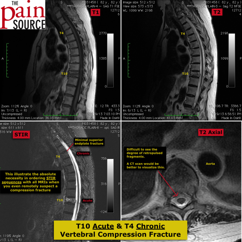
2) T10 Acute & T4 Chronic – Vertebral Compression Fractures
82 year old female that presented with pain at the lower thoracic region. This series of images shows the importance of getting STIR sequence images in addition to the regular MRI without contrast. Ordering with STIR does NOT cost more; it only takes a few more minutes for the radiology tech to perform.
Note that the small T4 superior end-plate fracture does NOT light up on STIR images, showing that it is chronic. On the other hand, the much more obvious T10 fracture is highly intense on the STIR images, which shows it is an acute process.
Update: This sweet old lady got better without the need for kyphoplasty or high dose narcotic medications.
